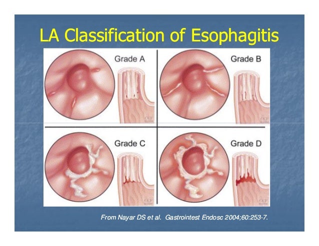


#La grade a relux esophagitis Patch
Superficial spreading carcinoma is a confluent patch or longer segment of poorly defined nodules. About 10% of patients with Barrett's esophagus demonstrate a “reticular pattern,” often adjacent to the distal aspect of a reflux-induced stricture.

Therefore, the “reticular pattern” of Barrett's esophagus mimics the reticular configuration of the areae gastricae of the stomach, though the tufts are smaller, about 1 mm in size. Barrett's esophagus has varying forms of columnar metaplasia arranged into tufts. Normal and abnormal columnar epithelium in the gastrointestinal tract is organized into tufts of mucosa, separated by shallow grooves. The clinical history of radiation is known. The esophagitis is confined to the radiation portal. The mucosa may be finely ulcerated or nodular in patients with acute radiation esophagitis. These nodules are usually separated by normal smooth mucosa. The nodules of glycogenic acanthosis, an aging variant, are more discrete, of variable size up to 3-4 mm, and are located in the upper and mid thoracic esophagus. Patients are usually symptomatic and many have gastroesophageal reflux or a hiatal hernia. The nodules in reflux esophagitis are poorly defined and located in the lower esophagus. Differential Diagnosis Finely Nodular Mucosa, Esophagus Nodularity of the contour of the folds in reflux esophagitis reflects mucosal nodularity seen in profile. The longitudinal folds of the esophagus are transient structures appearing when the esophagus collapses. Other findings of reflux esophagitis may be present, including thickened nodular folds (see Fig. Most patients have a hiatal hernia, and gastroesophageal reflux is seen at fluoroscopy. 1), termed “mucosal granularity” or “granular mucosa.” The nodules or granules of reflux esophagitis are confluent, located in the distal esophagus. Reflux esophagitis is typified by tiny, ill-defined elevations of the mucosal surface (see Fig. Patients are usually symptomatic, and many have gastroesophageal reflux or a hiatal hernia. Rubesin, in The Teaching Files: Gastrointestinal, 2010 DISCUSSION Definition/Background No malignant potential but may need endoscopy with biopsy to rule out cancer Single enlarged fold arising at GE junction □ Probably result and not cause of reflux – Seen in > 95% of patients with peptic stricture □ Transverse folds due to vertical scarring – Some may resemble Schatzki rings, but are generally thicker – Peptic stricture (1-4 cm length/0.2-2 cm width) □Ĭoncentric smooth-tapered narrowing of distal esophagus with proximal (upstream) dilatation □ Sacculations and pseudodiverticula may be seen – Thickened vertical or transverse folds (> 3 mm) ○ĭecreased distal esophageal distensibility with irregular, serrated contour (due to ulceration/edema/spasm) □ Single or multiple tiny collections of barium with surrounding mounds of edematous mucosa □ Decreased primary wave of peristalsis with increased tertiary contractions –įine nodular, granular, or discrete plaque-like defects (pseudomembranes) –


 0 kommentar(er)
